Ultrasound Procedure
29
Jun
2022
What is an Ultrasound?
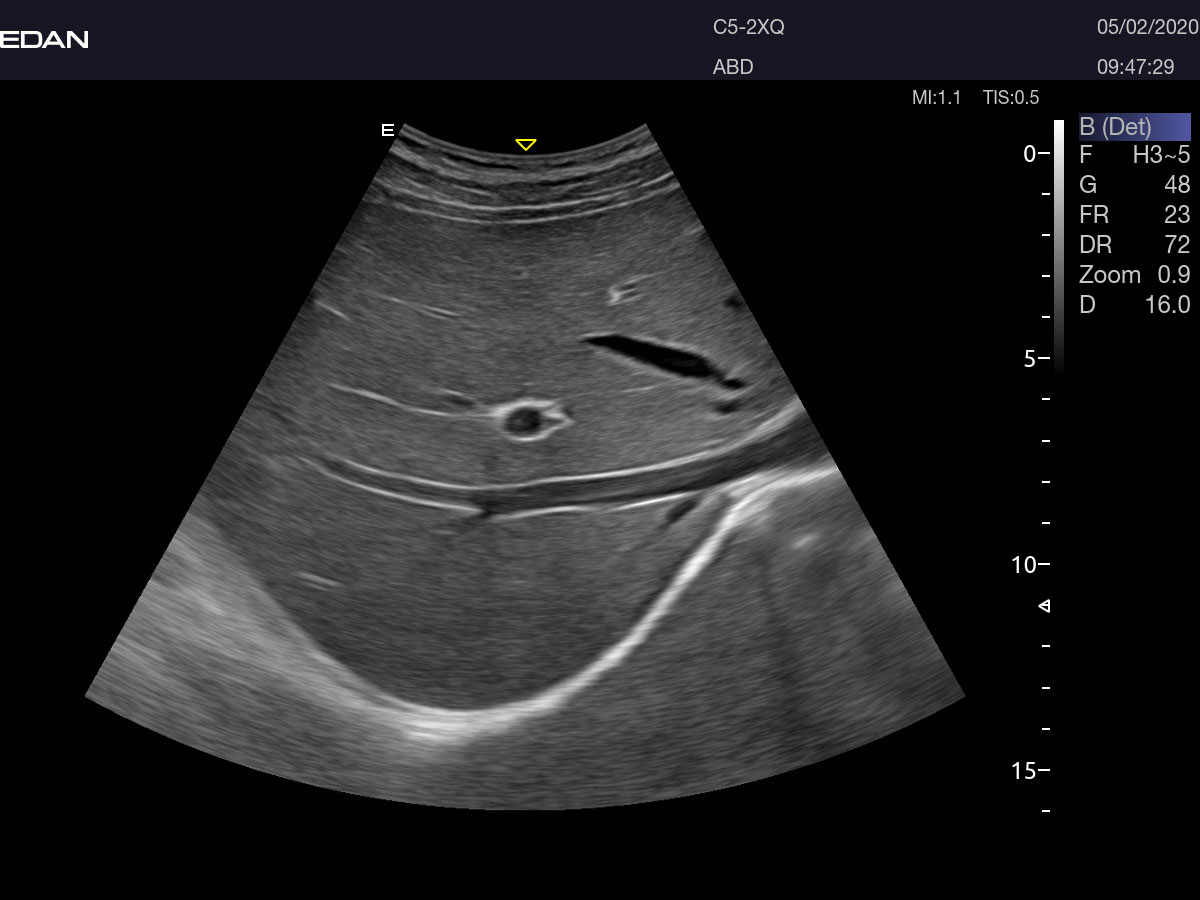
Diagnostic ultrasound, also known as sonography or diagnostic medical sonography, is an imaging method that uses sound waves to produce pictures of structures within your body. The images could provide valuable information for diagnosing and directing treatment for a variety of diseases and conditions.
Most ultrasound examinations are done using an ultrasound device outside your body, though some involve placing a tiny device inside your body.
Why it is done
Ultrasound is used for a number of reasons, including to:
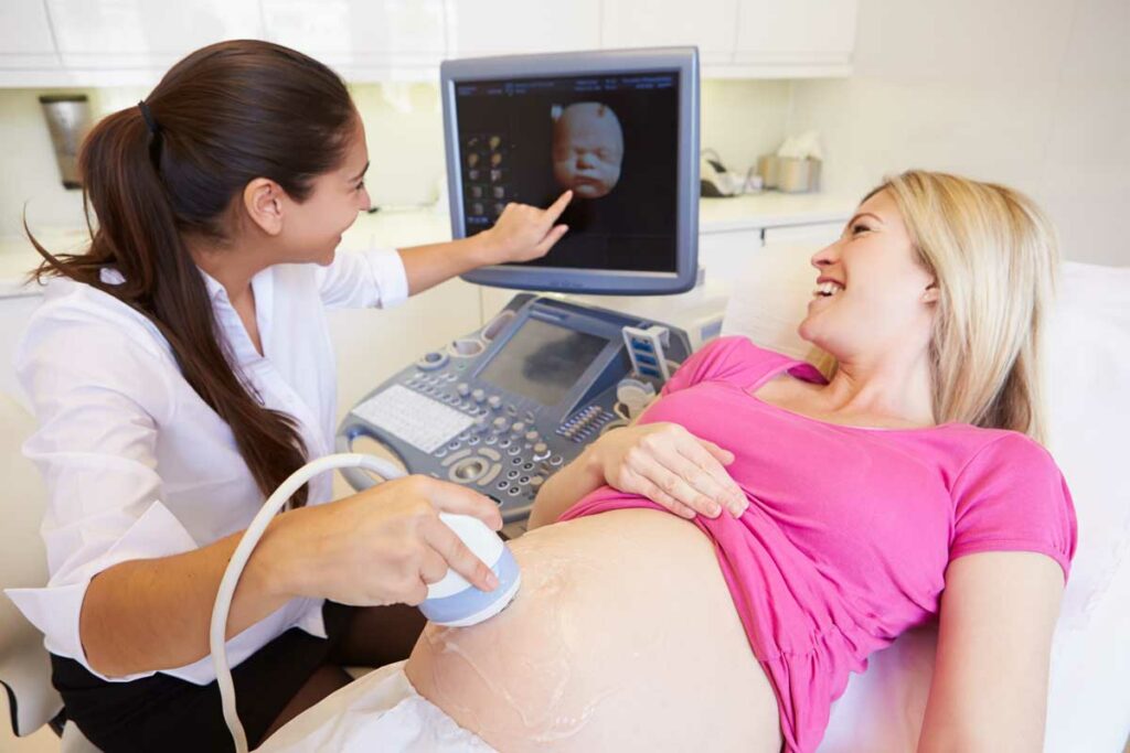
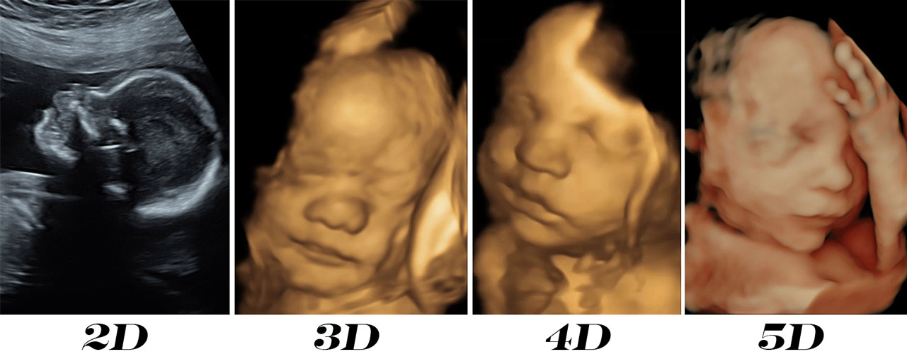
- Observe the uterus and ovaries during pregnancy and monitor the developing baby's health
- Diagnose gallbladder disease
- Evaluate blood flow
- Guide a needle for biopsy or tumor treatment
- Examine a breast lump
- Check the thyroid gland
- Find genital and prostate problems
- Assess joint inflammation (synovitis)
- Evaluate metabolic bone disease
Ultrasound Risks
Diagnostic ultrasound is a low-risk procedure that uses low-power sound waves. There aren’t any known risks.
Ultrasound is a valuable tool, but it has restrictions. Sound waves do not travel well through air or bone, so ultrasound is not effective at imaging body parts that have gas in them or are hidden by bone, like the lungs or head. Ultrasound might also be unable to see objects that are located very deep in the human body. To view these areas, your health care provider might order other imaging tests, such as CT or MRI scans or X-rays.
Ultrasound Preparation
Most ultrasound examinations require no preparation. However, there are some exceptions:
- For some scans, such as a gallbladder ultrasound, your care provider might ask that you not eat or drink for a certain period of time before the examination.
- Others, such as a pelvic ultrasound, might require a full bladder. Your doctor will let you know how much water you need to drink before the examination. Do not urinate until the examination is done.
- Young children might need additional preparation. When scheduling an ultrasound for yourself or your child, ask your doctor if there are any specific instructions you will need to follow.
Clothing and personal items
Wear loose clothing for your ultrasound appointment. You might be asked to remove jewelry during your ultrasound, so it is a good idea to leave any valuables at home.
What you can expect
Before the procedure
Before your ultrasound begins, you might be asked to do the following:
- Remove any jewelry from the region being examined.
- Remove or reposition all or a portion of your clothing.
- Change into a gown.
You will be prompted to lie down on the examination table.
During the procedure
The gel is applied to your skin over the region being examined. It helps prevent air pockets, which can block the sound waves that create the pictures. This safe, water-based gel is easy to remove from skin and, if needed, clothing.
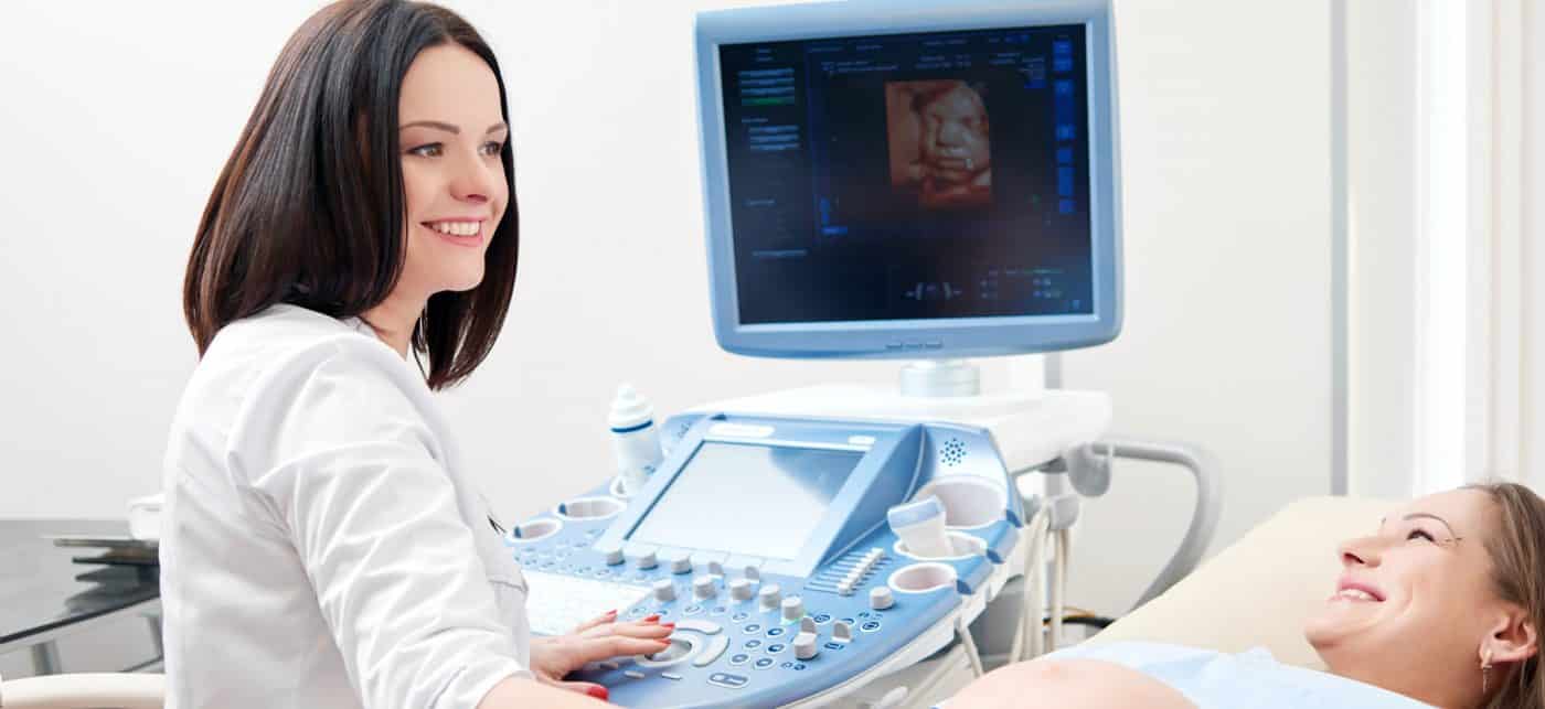
A trained technician (sonographer) presses a small, hand-held device (transducer) against the region being studied and moves it as needed to capture the images. The transducer sends sound waves into your body, collects the ones that bounce back, and sends them to a computer, which creates the pictures.
Sometimes, ultrasounds are performed inside your body. In this case, the transducer is connected to a probe that is inserted into a natural opening in your body. Examples include:
- Transesophageal echocardiogram - A transducer, inserted into the esophagus, obtains heart pictures. It is usually done while under sedation.
- Transrectal ultrasound - This test creates pictures of the prostate by placing a special transducer into the rectum.
- Transvaginal ultrasound - A special transducer is gradually inserted into the vagina to look at the uterus and ovaries.
Ultrasound is generally painless. However, you might experience mild discomfort as the sonographer guides the transducer over your body, particularly if you are required to have a full bladder, or inserts it into your body.
A typical ultrasound examination takes from thirty minutes to an hour.
Ultrasound Results
When your examination is complete, a doctor trained to interpret imaging studies (radiologist) analyzes the pictures and sends a report to your doctor. Your doctor will let you know about the results. You should be able to get back to normal activities immediately after an ultrasound.
When your examination is complete, a doctor trained to interpret imaging studies (radiologist) analyzes the pictures and sends a report to your doctor. Your doctor will let you know about the results. You should be able to get back to normal activities immediately after an ultrasound.
Hill Regional (HRH) Hospital is here to assist with all your medical needs with specialists and surgeons trained and experienced in the most advanced treatments. Our highly qualified doctors, nurses, and administrators are dedicated to caring for you with compassion in our state-of-the-art facilities.
Call us on 254-580-8500 to book an appointment with our specialist doctors.